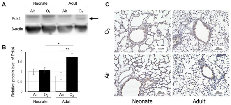
Fig 6. Expression levels of Pdk4 protein in the lungs quantified by Western blot (A,B) and immunohistochemical localization of Pdk4 protein in the lung (C).
(A) The arrow indicates a band of Pdk4 protein. (B) The vertical axis shows the relative expression of Pdk4 protein. Data are shown as means ± SEM. Comparisons between groups were performed using a t test. * p < 0.05; ** p < 0.01. Air: normoxia; O2: hyperoxia; SEM: Standard error of the mean. (C) Immunostaining was observed in the epithelium of terminal bronchiole in all four groups (brown). Scale bar = 100 μm.

