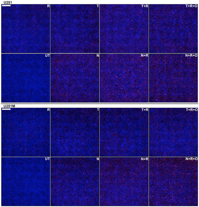
Fig 6. Quantitative ICC analysis of irreparable DNA damage in U251/U251M GB cells.
Cells were seeded at high densities (50,000 cells/cm2) and either left untreated (UT) or treated for five consecutive days with either 10 μM TMZ (T) or NEO212 (N) or 2 Gy (R) alone or combinations without (T+R or N+R) or with (T+R+O or N+R+O) Olaparib (O). The cells were probed with a γH2AX antibody and an AF647-labeled secondary and nuclei were counterstained with DAPI. Persistent γH2AX foci (red) were digitally counted relative to the total number of cell nuclei (blue). Each panel is data dense and represents a composite of 36 fields in total (i.e., a square of about 3x3 mm) captured on a widefield microscopy instrument and digitally stitched together. Scale bar is 500 μm (upper left corner).

