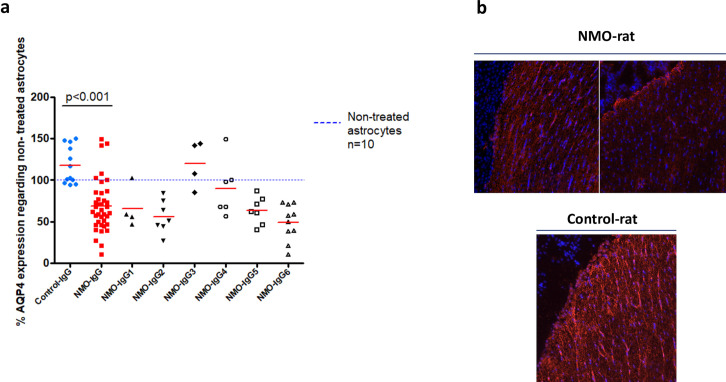
Fig 1. Aquaporin-4 expression in astrocyte culture and rat brain tissue.
(a) Expression of AQP4 in astrocytes after 24 hours of treatment with NMO-IgG1-6 (n = 39) compared to treatment with Control-IgG (n = 12). Dash blue line represents non-treated astrocytes (Wb). (b) Example of AQP4 expression in the periventricular regions of rat brains after being treated with NMO-IgG (NMO-rat) compared to Control-IgG (Control-rat) (immunohistochemistry).

