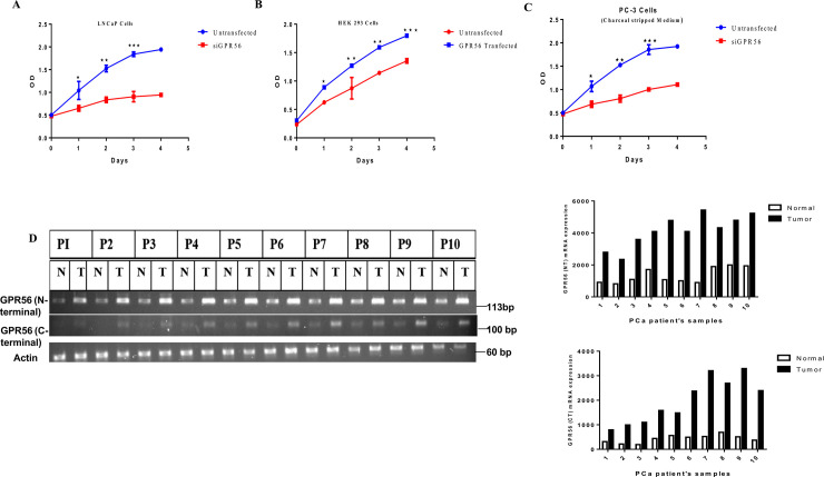
Fig 9. GPR56 expression in tumor samples.
A B & C) MTT assay in LNCaP, HEK 293 and PC3 cells. Cell proliferation in LNCaP cells transfected with GPR56-siRNA. Cell proliferation in LNCaP cells and cells transfected with siGPR56 were examined at 24 h, 48 h, 72 h or 96 h after seeding, using MTT assay. The data represents the mean ±S.D. of three independent experiments. Values are mean ±S.D. from three independent experiments. * p< 0.01, ** p<0.001, *** p<0.0001, two-way anova test B) Cell proliferation in non-transfected and GPR56 transfected HEK cells was examined 1–4 days after seeding using MTT assay. The data represents the mean ±S.D. of three independent experiments. * p< 0.01, ** p<0.001, *** p<0.0001, two-way anova test. Cell proliferation in PC3 cells transfected with GPR56-siRNA. Cell proliferation in PC3 cells and cells transfected with siGPR56 were examined at 24 h, 48 h, 72 h or 96 h after seeding, using MTT assay. The data represents the mean ±S.D. of three independent experiments. Values are mean ±S.D. from three independent experiments. * p< 0.01, ** p<0.001, *** p<0.0001, two-way anova test D) Representative RT-PCR analysis of GPR56 in matched normal (N) vs prostate tumor (T) tissue from individual patient’s samples (P1-P10) (P11-P25) patient’s samples. RNA was isolated from prostate tumor (T) and matched normal (N) tissue from individual patients and reverse transcribed using gene specific primers for GPR56 (as described in materials and methods). The amplified products were resolved on 2% agarose gel. The bands corresponding to GPR56 (N- terminal)-143 bp and GPR56 (C-terminal)-110 bp were observed. Beta actin was used as control. Quantification of band densities for N-terminal or the C- terminal region of the GPR56 mRNA transcript in patient’s tissue samples has been shown (using Image J).

