
Fig. 1.
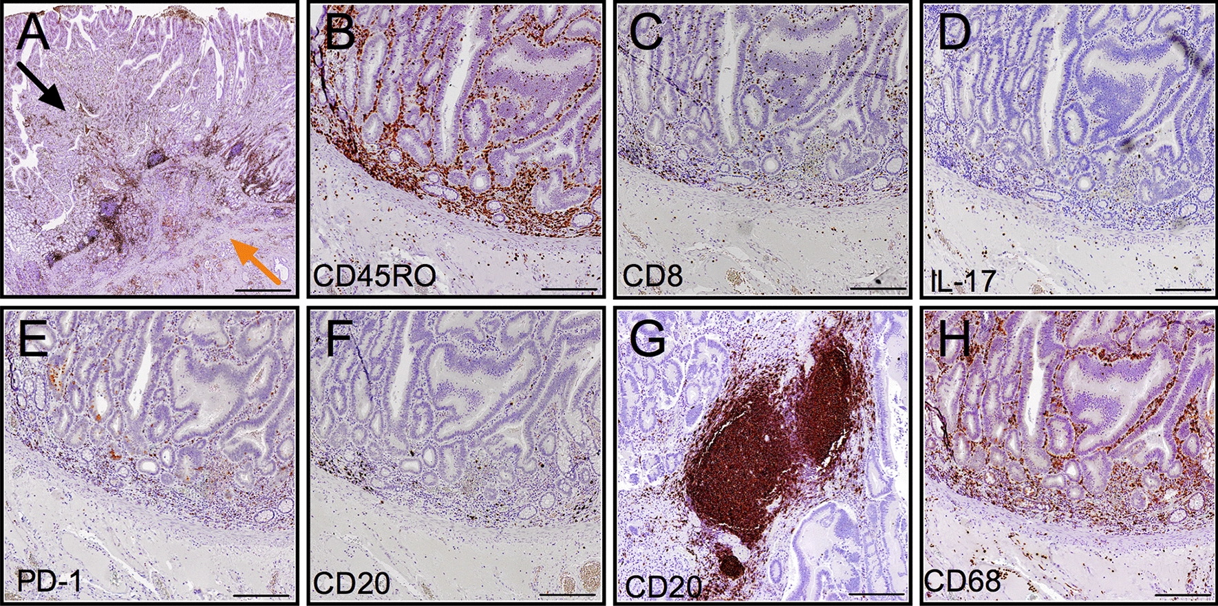
Immunohistochemical evaluation of immune cells in human duodenal cancer. a Representative picture of a human duodenal adenocarcinoma specimen; the invasive front (orange arrow) and tumor core regions (black arrow) are indicated. b–h Representative pictures of CD45RO-TILs (b), CD8-TILs (c), IL17+ cells (d), PD1-TILs (e), CD20-TILs (f), CD68-TAM (h) at the invasive margin and CD20-TLT (g) in the tumor stroma. Scale bars = 500 μm (a), 200 μm (b–h). TILs: tumor-infiltrating lymphocytes; TLT: tertiary lymphoid tissue; TAM: tumor-associated macrophages