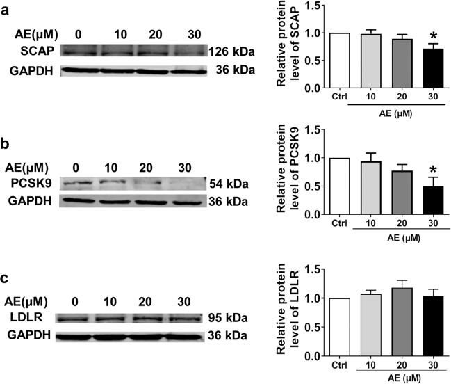
Fig. 5.
Effects of AE at different concentrations ranging from 10 to 30 μM on the protein levels of SCAP, PCSK9, and LDLR in HepG2 cells. a Western blot band of SCAP and statistical results. b Western blot bands of PCSK9 and statistical results. c Western blot bands of LDLR and statistical results. *P < 0.05 vs. Ctrl, n = 6

