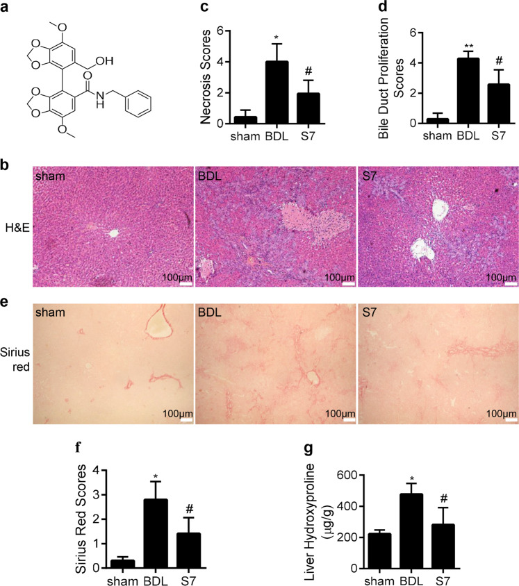
Fig. 1. IMB-S7 ameliorates BDL-induced liver injury in rats.
a Chemical structure of IMB-S7. b Liver samples collected from three groups (sham, BDL, and IMB-S7) were examined by H&E staining, and representative pathological images are shown (×40, n = 6 per group). c, d Quantitative assessment of liver necrosis and bile duct proliferation. *P < 0.05, **P < 0.01 vs the sham group; #P < 0.05 vs the BDL group. e, f Liver samples collected from three groups (sham, BDL, and IMB-S7) were examined by Sirius red staining, representative images of Sirius red staining (×40, n = 6 per group) and positive staining scores. g Hydroxyproline content in liver samples was determined by a hydroxyproline assay kit. Data are expressed as the mean ± SD of three independent experiments, *P < 0.05 vs the sham group; #P < 0.05 vs the BDL group.

