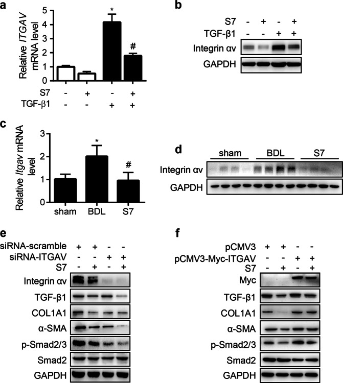
Fig. 3. IMB-S7 suppresses the expression of integrin αv.
a, b LX2 cells were starved for 24 h and then treated with TGF-β1 (2 ng/mL) and IMB-S7 (0.2 mM) for an additional 24 h. Real-time PCR (a) and Western blot analysis (b) for integrin αv, *P < 0.05 vs the left column; #P < 0.05 vs the TGF-β1 column. Real-time PCR (c) and Western blot analysis (d) for integrin αv in liver samples of the BDL rat model (n = 6 per group), *P < 0.05 vs the sham group; #P < 0.05 vs the BDL group. LX2 cells were plated and cultured in six-well plates overnight, followed by siRNA-ITGAV (e) or pCMV-Myc-ITGAV (f) for 48 h, followed by IMB-S7 (0.2 mM) for an additional 24 h. Western blot analysis for the indicated proteins.

