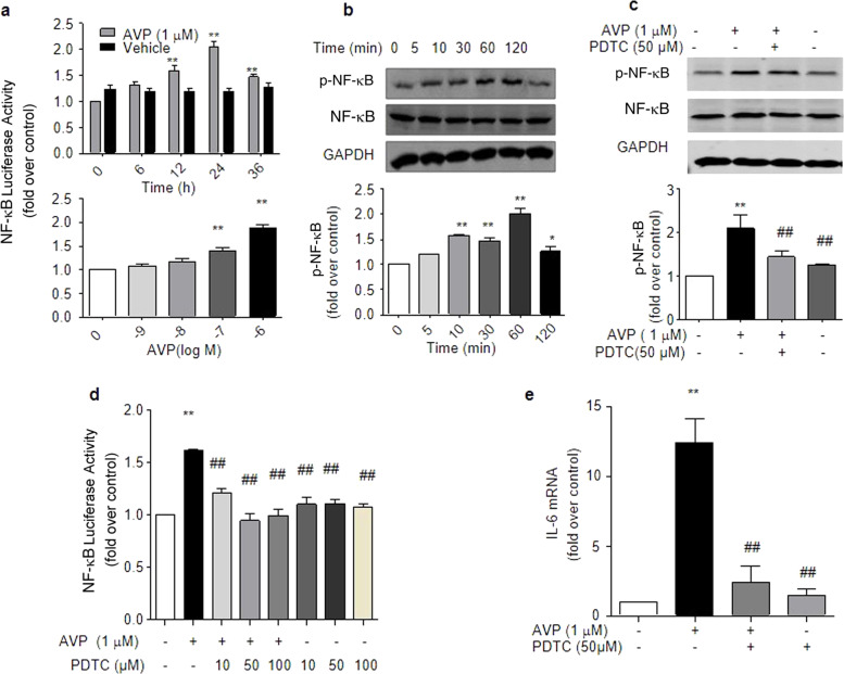
Fig. 4.
AVP induced IL-6 production via NF-κB in ARCFs. a AVP evoked the activation of NF-κB in a dose- and time-dependent manner. Cells transfected with the NF-κB luciferase reporter plasmid were incubated with 10−6 M AVP for 0–36 h or with AVP for 24 h. Luciferase activation was measured as described in the kit manual. The data are the averages of three separate experiments. *P < 0.05, **P < 0.01 vs. control. b AVP induced the phosphorylation of NF-κB in a time-dependent manner. Starved cells were stimulated with 10−6 M AVP for 0–120 min. Cellular lysates were collected to measure the phosphorylation of NF-κB using an anti-phosphorylated NF-κB antibody, as described in the Methods. The upper panel is a representative blot, and the lower panel shows the average data of three separate experiments. #P < 0.05 for time-course (repeated two-way ANOVA, n = 3). *P < 0.05, **P < 0.01 vs. control (one-way ANOVA). c, d The inhibition of NF-κB by PDTC abolished the AVP-induced phosphorylation and activation of NF-κB-luciferase. Starved cells were pretreated with 50 µM PDTC for 1 h and further incubated with 1.0 µM AVP for 1 h. Cellular lysates were collected to measure NF-κB phosphorylation using an anti-phosphorylated NF-κB antibody, as described in the Methods. The activation of luciferase in cells treated with 1.0 µM of AVP for 24 h was assayed as described in the Methods. e The inhibition of NF-κB by PDTC suppressed the AVP-induced levels of IL-6 mRNA. Starved cells were pretreated with 50 µM PDTC for 1 h and further incubated with 1.0 µM AVP for 24 h. The supernatants were harvested to measure IL-6 mRNA. The average data are from four separate experiments. *P < 0.05, **P < 0.01 vs. control, ##P < 0.01 vs. AVP alone. Notably, PDTC alone had no effect on the AVP-induced phosphorylation and activation of NF-κB or on IL-6 mRNA levels

