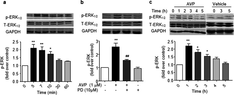
Fig. 6.
AVP induces the phosphorylation of ERK1/2 via a PD98059-sensitive pathway in cultured adult cardiac fibroblasts (a, b) and rat heats (c). a, b Treatment with 0.1 µM AVP evoked the phosphorylation of ERK1/2 in a time-dependent manner (a). These effects were inhibited by 10 µM PD98059 (b). The upper panels in a and b are representative images and the lower panels show the data expressed as the means ± SEM of three separate experiments. P < 0.05 for time-course (repeated two-way ANOVA), *P < 0.05, **P < 0.01 vs. control. c AVP evoked phosphorylation of ERK1/2 in rat hearts. After AVP (0.5 U/kg) was injected into the tail vein for 0–5 h, the left ventricles were harvested to obtain ventricular lysates to detect phosphorylated and total ERK1/2. The upper panels are representative images, and the lower panels show the data expressed as the means ± SEM of three separate experiments. *P < 0.05, **P < 0.01 vs. control

