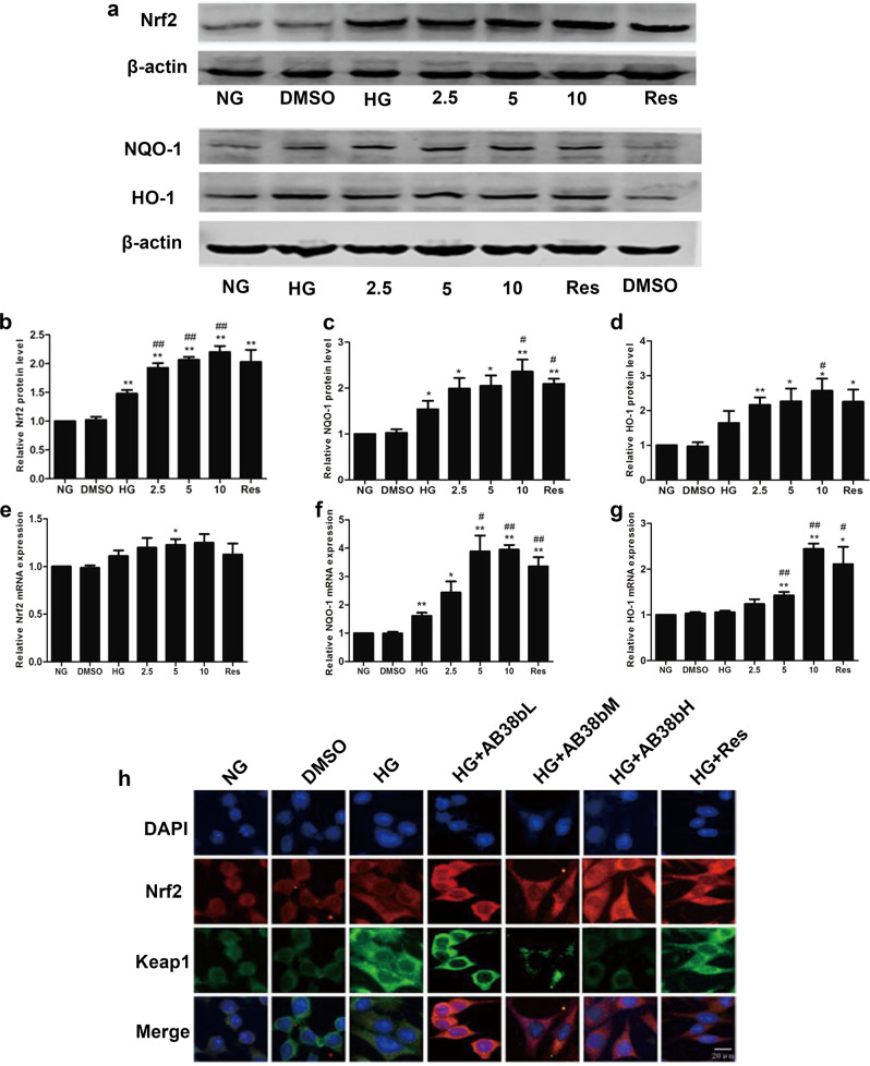
Fig. 7.
Effects of AB38b on Nrf2 signaling in high glucose-induced mesangial cells. a Western blot analysis of Nrf2 (upper panel), NQO-1, and HO-1 (bottom panel) in the different groups of mesangial cells. b–d The densitometric analysis of the Western blots. The Nrf2 b, NQO-1 c, and HO-1 d signals were normalized to the β-actin signals for the same samples to determine the fold-change relative to the control, the expression level of which was set as 1. e–g The relative mRNA expression levels of Nrf2 e, NQO-1 f, and HO-1 g in the different groups of mesangial cells. h Confocal immunofluorescence images showing the colocalization of Nrf2 and Keap1 in SV40 cells exposed to HG conditions and treated with various concentrations of AB38b. The data are expressed as the mean ± SD, n = 3. *P < 0.05, **P < 0.01 vs. NG; #P < 0.05, ##P < 0.01 vs. HG

