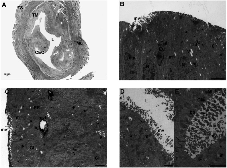
Fig. 1.
Control group. A) Representative LM picture of a transversal section of mouse ampulla showing the three layers of tunica mucosa (TM), muscularis (TMu) and serosa (TS). The TM appears regularly folded and characterized by adjacent columnar epithelial cells (CEC) with evident nuclei (*). In the tunica muscularis (TMu), the two well-defined outer and inner layers are evident. The former is organized in individual smooth muscle fiber bundles longitudinally oriented while the latter is organized in circular fibers. L: lumen (LM. Mag: 20 ×. Bar: 5 μm). B) Ultrastructure of the columnar epithelium of TM, showing roundish-to-amoeboid shaped nuclei (N) delimited by a continuous nuclear membrane (nm). Heterochromatin (He) was clustered in clumps or located as marginal patches under the nuclear envelope among the dispersed euchromatin (Eu). The cytoplasm contains numerous round or elongated mitochondria (m), with electron-dense lamellar cristae or electron-pale content; lipid droplets (ld), short and long microvilli (mv), a few electron-negative vacuoles (V) and secretory granules with a slightly electron-dense content (asterisks). The junctional complexes between neighboring epithelial cells appear well-formed and constituted from the lumen by zonulae occludens (arrow) followed by zonulae adherens (arrowhead) [transmission electron microscopy (TEM). Bar: 2 μm]. C) CECs of TM showing a large multivesicular body (mvb), an electron-dense secondary lysosome (Ly), tubular elements of the endoplasmic reticulum (ER), well-defined nuclei (N) and mitochondria (m). V: vacuoles; mv: microvilli; Gl: glycogen granules; asterisk: secretory granules (TEM. Bar: 1 μm). D) The luminal side (L) of TM shows numerous short or long microvilli (mv). m: mitochondria; arrows: zonulae occludens; arrowhead: zonula adherens (TEM. Bar: 1 μm). E) High magnification of motile cilia (Ci) showing an evident axoneme of nine peripheral doublet microtubules, surrounding a central complex with two central microtubules and the central sheath (9 + 2 arrangement). Gl: glycogen granule; arrows: zonula occludens (TEM. Bar: 2 μm).

