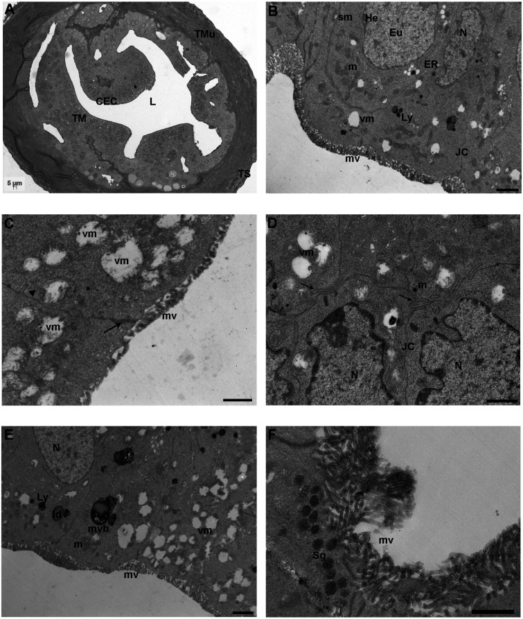
Fig. 3.
8 round (R) group. A) Representative image of a semithin section of the ampullar region of mouse FTs showing the highly folded tunica mucosa (TM) with big nuclei (asterisk) and the well-defined two layers of tunica muscularis (TMu) and the tunica serosa (TS). CECs: columnar epithelial cells, L: lumen (LM. Mag: 20 ×. Bar: 5 μm). B) Micrograph of the tunica mucosa showing nuclei (N), round/elongated electron-dense mitochondria (m), endoplasmic reticulum (ER) tubules/networks, secondary lysosomes (Ly), microvilli (mv) and junctional complexes (JC). Eu: euchromatin; He: heterochromatin, sm: swollen mitochondria; vm: vacuolated mitochondria [transmission electron microscopy (TEM). Bar: 1 μm]. C) Detail of damaged mitochondria showing numerous mitophagic vacuoles (vm) characterized by partial or complete cristolysis and peripheric accumulation of the mitochondrial remains. mv: microvilli; arrow: zonula occludens; arrowhead: zonulae adherens (TEM. Bar: 0.8 μm). D) Epithelial cells showing interdigitated cell contacts (arrows). The cytoplasm contains irregularly shaped nuclei (N) and numerous vacuolated mitochondria (vm). JC: junctional complexes (TEM. Bar: 1 μm). E) Cytoplasmic content of columnar epithelial cells showing numerous mitophagic vacuoles (vm) in proximity to multivesicular bodies (mvb) and lysosomes (Ly). ld: lipid droplet; mv: microvilli; m: mitochondria; N: nucleus (TEM. Bar: 1 μm). F) High magnification of non-ciliated cells rich in electron-dense secretory granules (Sg) with homogeneous content and numerous long and thin microvilli (TEM. Bar: 1 μm).

