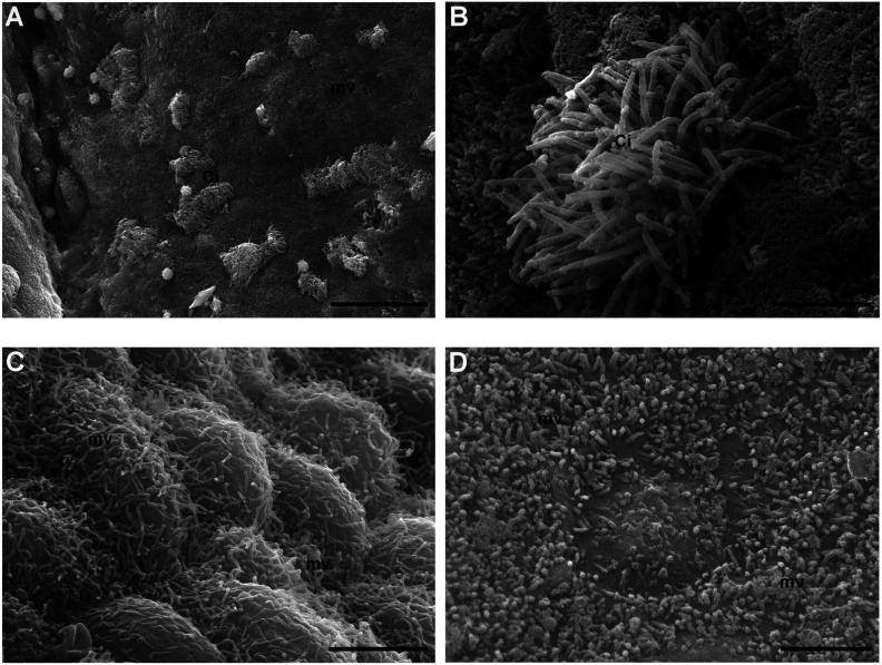
Fig. 4.
Surface analysis by scanning electron microscopy (SEM). A–B) Control group. A) Numerous tufts of cilia (Ci) surrounded by short microvilli (mv) protrude into the lumen of the ampullar epithelium (SEM. Bar: 30 μm). B) High magnification of a well-preserved tuft of cilia (Ci) (SEM. Bar: 3 μm). C–D) 4 round (R) group. C) Numerous, long and homogenously distributed microvilli (mv) protrude from the globoid surface of epithelial cells (SEM. Bar: 3 μm). D) Detail of a heterogeneous carpet of short microvilli (mv) (SEM. Bar: 2.5 μm).

