Figure 5.
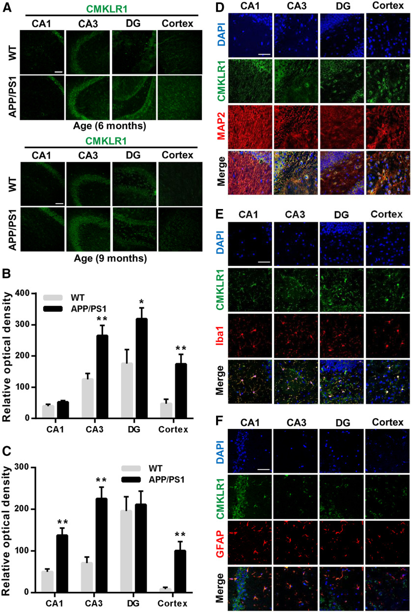
Upregulation of CMKLR1 and colocalization with MAP2, Iba1, and GFAP in the brain of APP/PS1 mice. A, Representative immunofluorescence images of CMKLR1 (green) in CA1, CA3, and DG regions of the hippocampus and in the cortex of WT and APP/PS1 mice aged 6 months and 9 months. Scale bar, 50 µm. Quantification of the fluorescence intensity of CMKLR1 in each of the regions, showing that CMKLR1 was significantly increased in the hippocampus and the cortex of APP/PS1 mice at the age of 6 months (B) and 9 months (C). Data are mean ± SEM, with at least 3 mice in each group. *p < 0.05; **p < 0.01; compared with WT mice. D–F, Colocalization of CMKLR1 (green) with neurons (anti-MAP2, red, D), microglia (anti-Iba1, red, E), and astrocytes (anti-GFAP, red, F) in the hippocampus and the cortex of APP/PS1 mice aged 9 months. Scale bar, 50 μm.

