Figure 9.
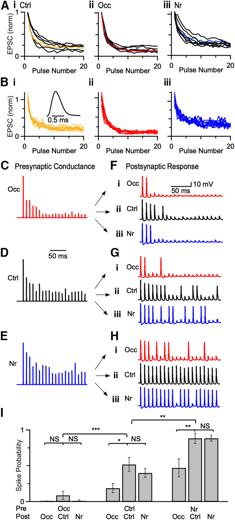
Presynaptic versus postsynaptic contributions to spike fidelity. A, Average EPSC amplitudes from voltage-clamp experiments in response to a 20-pulse train at 100 Hz from (Ai) 6 control, (Aii) 7 occluded, and (Aiii) 8 noise-reared endbulbs. Data were normalized to EPSC1. Cells with intermediate levels of synaptic depression were selected as representative cells for each acoustic condition (colored lines). B, Individual trials from the representative endbulbs from different acoustic conditions in A (Ctrl: P21, 12 trials; Occ: P22, 9 trials; Nr: P23, 11 trials). These individual amplitudes were convolved with a unit conductance (Bi, inset) and scaled by the threshold of the cell. C–E, Example AMPA conductances derived from data in B. F–H, Examples of two-electrode dynamic-clamp recordings from occluded (i), control (ii), and noise-reared (iii) BCs after applying representative conductances in C–E. I, Spike fidelity for presynaptic conductances applied to postsynaptic BCs from three acoustic conditions. Firing probability was quantified from the last 10 stimuli of 20 pulse trains, when conductance amplitudes reach steady state. Presynaptic changes affected spike fidelity after occlusion and noise-rearing, while postsynaptic changes affected spiking primarily after occlusion. *p < 0.05, **p < 0.01, ***p < 0.001, NS = not significant (p > 0.05).

