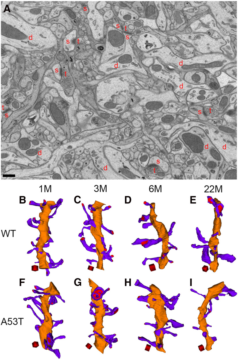
Figure 1.
3D reconstruction of dendrites and spines from WT and A53T-BAC-SNCA mice at different ages. A, Membrane contours of dendrites (d), spines (s), and presynaptic terminals (t) can be clearly observed in the FIB/SEM image from a 1-month-old WT mouse. Mitochondria and other organelles are also clearly visible. Scale bar: 500 nm. B–I, 3D reconstruction of dendrites (orange) and spines (violet) from WT mice at 1 (B), 3 (C), 6 (D), and 22 (E) months of age and from A53T-BAC-SNCA mice at 1 (F), 3 (G), 6 (H), and 22 (I) months of age. Red regions in the spine heads indicate postsynaptic densities. Scale cubes: 0.5 µm on each side.

