Figure 4.

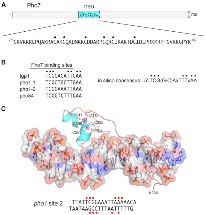
Transcription factor Pho7 recognizes a UAS element in the PHO mRNA promoters. (A) The 738-aa Pho7 polypeptide is depicted as a horizontal bar. The internal DNA-binding domain containing the Cys6•Zn2 module is colored cyan. The primary structure of the minimal DBD is shown below the cartoon, with the Zn-binding cysteines indicated by dots. (B) The nucleobase sequences at the experimentally determined Pho7 binding sites in the indicated PHO mRNA promoters are aligned. Conserved positions are indicated by black dots. The Pho7 binding site consensus sequence identified by in silico analysis of genome-wide Pho7 ChIP-seq data is shown on the right. (C) Structure of Pho7 DBD bound to pho1 site 2 DNA. The Pho7–DNA complex is shown with the DNA depicted as a stick model, with an overlying transparent surface model to highlight the major and minor grooves. The Pho7 protein is rendered as a cartoon trace with cyan α-helices. The two zinc atoms are shown as green spheres and the six zinc-binding cysteines are shown as stick models and labeled. Anomalous difference density for the zinc atoms, contoured at 5σ, is shown in red mesh. The primary structure of the pho1 site 2 DNA ligand is shown at bottom. Red dots indicate DNA nucleobases that are contacted directly by Pho7 amino acids.
