Figure 4.
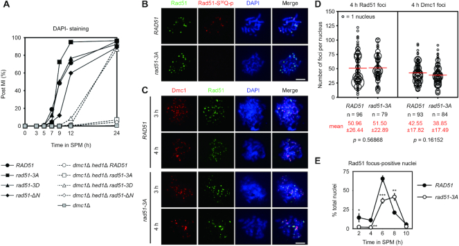
Rad51-NTD and its phosphorylation are indispensable for Rad51-only meiotic recombination during dmc1Δ hed1Δ meiosis. (A) Meiotic progression was monitored by DAPI (4′,6-diamidino-2-phenylindole)-staining of nuclei. Cells harvested from SPM having two or four nuclei (as determined by fluorescence microscopy) were assessed as having completed meiosis I (MI), and the percentage of such cells over total cells counted (n = 200) per time-point was plotted. (B, C) Cytology. Representative images of meiotic nuclear surface spreading experiments using guinea pig anti-Rad51 (green) and anti-phosphorylated Rad51-S30Q (red) or anti-Dmc1 (red) antisera, respectively. Meiotic chromosomes were stained with DAPI (blue) at indicated sporulation time-points. Scale bars, 5 μm. (D) Quantification of the numbers of Rad51 and Dmc1 foci in the WT and rad51-3A strains. The numbers of foci in each foci-positive chromosome spread (with more than five foci) were counted and plotted as shown. The sizes of circles are proportional to the numbers of nuclei with a given number of foci. The mean number of foci per nucleus is shown in red (bottom), and also as a red bar in the graph. Standard deviations of numbers of foci are shown in parentheses. n represents the number of nuclei analyzed for foci-counting. The P values were calculated using a two-tailed Mann–Whitney's U-test. (E) Kinetics of Rad51 foci in meiosis of WT and rad51-3A strains. For each replicate of experiments (n = 3), Rad51-foci-positive nuclei (with more than five foci) were examined in 60 chromosome spreads. Error bars indicate standard deviation between experiments. Asterisks indicate significant difference between WT and rad51-3A strains, with P values calculated using a two-tailed t-test (*P value < 0.05; **P value < 0.01 and ***P value < 0.001).

