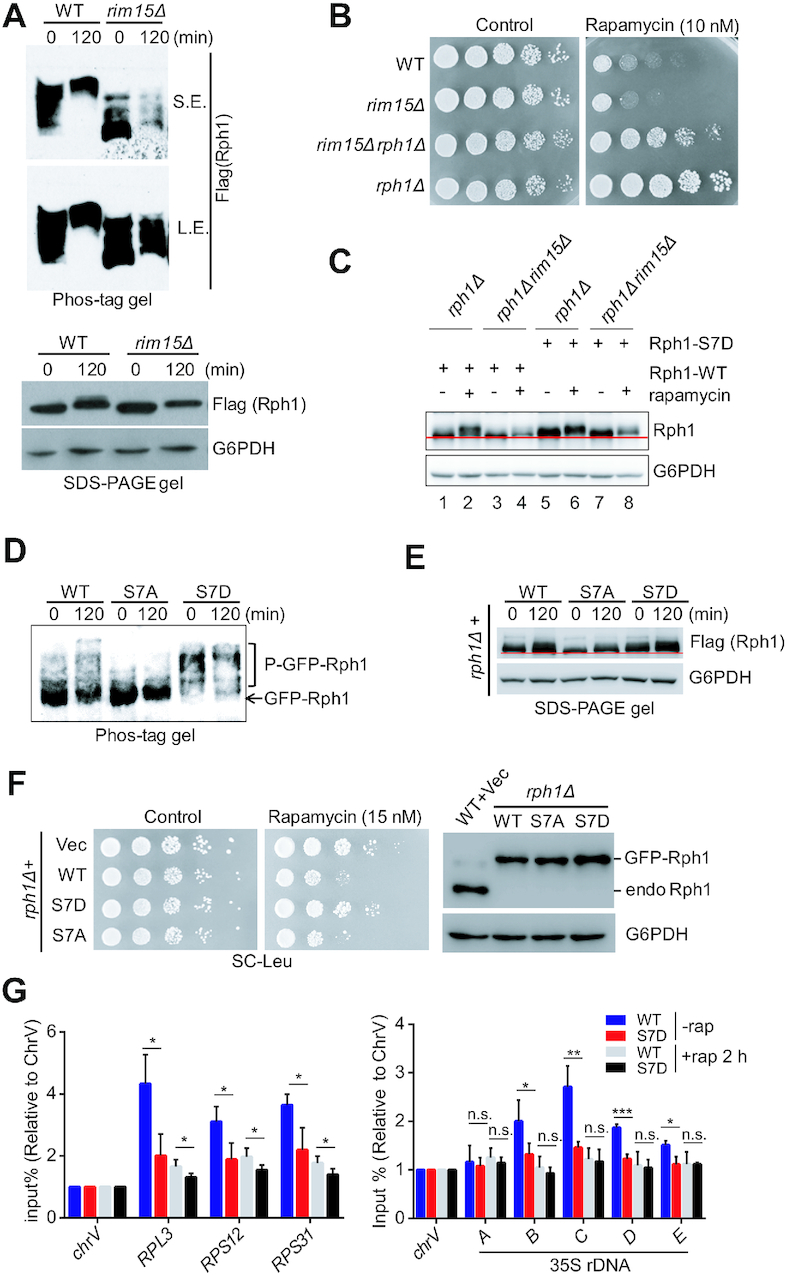
Figure 6.

Rim15-dependent Rph1 phosphorylation alleviates cellular sensitivity to rapamycin stress. (A) The phosphorylation states of Rph1-Flag in indicated strains upon rapamycin treatment were examined using phos-tag gel (top panel) or SDS-PAGE gel (bottom panel). (B) The indicated strains were spotted on YPD plates with or without rapamycin. (C) Mobility shift of the indicated cells bearing either WT or S7D Rph1 under rapamycin treatment was examined using regular SDS-PAGE gel. (D andE) The phosphorylation states of GFP-Rph1 or Flag-Rph1 bearing either the WT or mutations driven by endogenous RPH1 promoter upon rapamycin treatment were examined using a phos-tag gel (panel D) or SDS-PAGE gel (panel E), respectively. (F) Rapamycin sensitivity of various Rph1 constructs were monitored by a spot assay (left panel), and protein levels of GFP-Rph1 in the indicated cells were examined by immunoblotting (right panel). (G) ChIP-qPCR analysis showed relative abundance of WT or S7D Rph1 in different RP gene loci (left panel) and different regions of 35S rDNA (right panel) with or without rapamycin treatment. qPCR data are represented as mean ± SD from at least three biological replicates. t-test, ∗P < 0.05; ∗∗P < 0.01; ***P < 0.001, n.s. ‘not significant’
