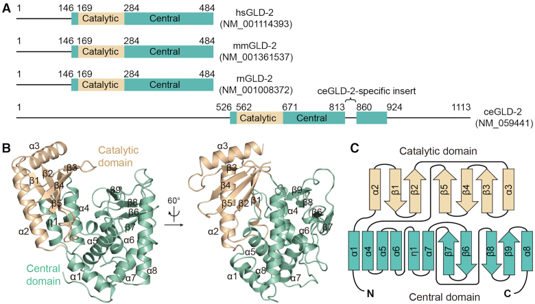
Figure 1.
Overall structure of GLD-2. (A) Schematic representation showing the domain organization of mammalian and Caenorhabditiselegans GLD-2 homologs. Borders of the domains are indicated by residue numbers. hs, Homo sapiens; mm, Mus musculus; rn, Rattus norvegicus; ce, Caenorhabditis elegans. (B) Cartoon representation of rnGLD-2, colored as in A. The identity of each helix and β-strand are indicated. (C) The topology diagram of rnGLD-2. Secondary structural elements are not drawn to scale. Elements of rnGLD-2 are named and colored as in B.

