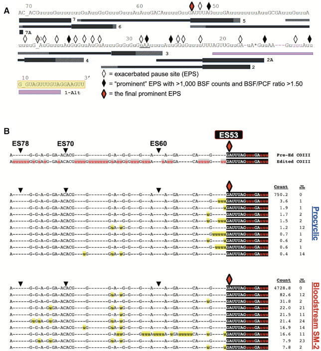
Figure 7.
Statistical and bioinformatics analysis of COIII mRNA intermediates at the final prominent EPS in PCF and BSF SM-2 cells. (A) Schematic illustrating the positions of exacerbated pause sites (EPSs) in BSF COIII mRNA in relation to the position of guide RNAs (gRNAs). Symbols as in Figure 5. The red diamond represents the final prominent EPS at ES53. (B) Sequence alignments of the 10 most abundant junction sequences at ES53 in COIII mRNA of PCF and BSF SM-2 cells. Symbols and shading as in Figure 5.

