Figure 1.

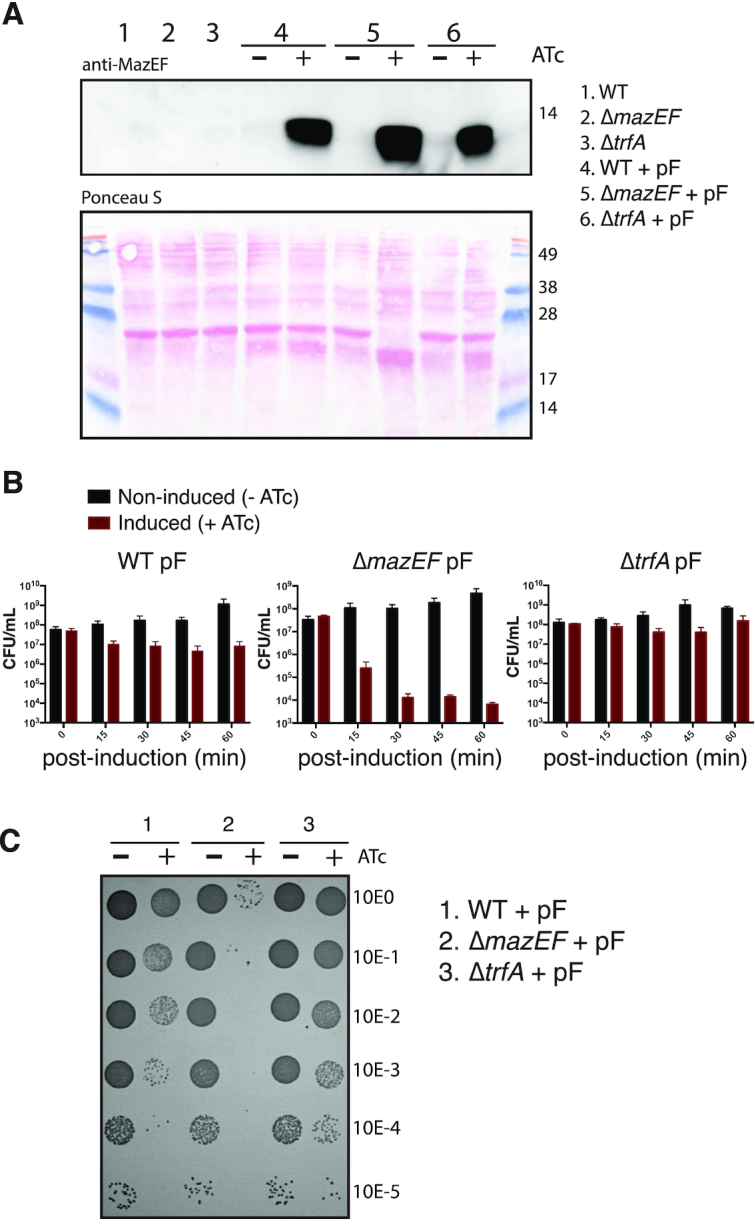
(A) Western blot analysis of MazF protein produced in S. aureus strains with or without the mazF overexpression plasmid (pF). After mazF gene induction with anhydrotetracycline (+ATc) or in non-induced cells (−ATc), total soluble protein extracts from S. aureus strains were loaded in SDS 16.5% polyacrylamide gels and MazF protein (13.4 kDa) was detected using a rabbit-polyclonal anti-MazEF antibody (top) and Ponceau S staining (bottom). A Coomassie Brilliant Blue stain on same samples loaded above is shown in Supplementary Figure S2. (B) Effect of mazF overexpression on S. aureus growth. Colony forming units (CFU) counts at five time points (between 0 and 60 min) after mazF-induced (+ATc) or uninduced (−ATc) S. aureus strains. Data are represented as mean ± SD of three independent experiments. (C) Representative image showing the spot (10 μl) serial dilutions of bacterial cultures on agar plates, after 60 min of mazF-induced (+ATc) and uninduced (−ATc) S. aureus strains. Serial dilutions are indicated at the right margin. CFU counts of S. aureus carrying an empty vector with and without ATc is shown in Supplementary Figure S3.
