Figure 2.

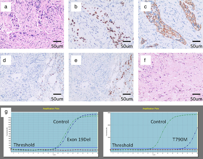
Pathology and amplification refractory mutation system‐polymerase chain reaction (ARMS‐PCR). (a) At the time of diagnosis. Hematoxylin and eosin (HE) staining showed neoplastic cells with morphological characteristics of non‐small cell lung cancer (NSCLC). (b–e) At the time of diagnosis. (b) Immunohistochemistry revealed diffuse expression of P40; (c) CK (5/6); (d) negative expression of TTF‐1; and (e) Napsin A, which led to the diagnosis of lung squamous cell carcinoma. (f) At the time of surgery. HE staining showed a necrotic area of former cancer tissue, with no residual viable cancer cells. (g) At the time of diagnosis. ARMS‐PCR showed coexistence of epidermal growth factor receptor (EGFR) exon 19Del and T790M (magnification: 200x; scale bar: 50 μm).
