
Figure 1.
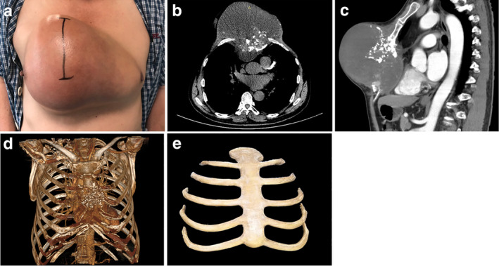
(a) A 59‐year‐old male patient presented with a large chest tumor located throughout the anterior chest wall. (b) Chest computed tomography (CT) showed the destruction of the sternum and large soft tissue masses. The right pectoralis major muscle was invaded by the tumor. (c) Sagittal chest CT showed a large mass had compressed the pericardium. (d) Mimics software was used to perform a concrete model of tumor destruction. (e) The 3DP PEEK implant weighed 206 g with bending strength at 141 ± 7 MPa and tensile strength at 89 ± 3 MPa, elastic modulus at 2.8 ± 1.5 GPa.