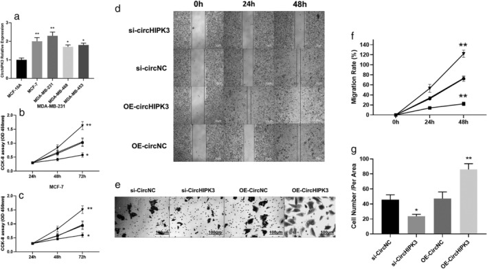
Figure 2.

(a) The expressions of CirCHIPK3 in normal and breast cancer cells. (b) MDA‐MB‐231 cell proliferation was detected by MTT assay following si‐CirCHIPK3 and OE‐ CirCHIPK3 (*P < 0.05,**P < 0.01) ( ) si‐CircHIPK3, (
 ) si‐CircNC, (
 ) OE‐CircNC, (
 ) OE‐CircHIPK3. (c) MCF‐7 cell proliferation was detected by MTT assay following si‐CirCHIPK3 and OE‐CirCHIPK3 (*P < 0.05,**P < 0.01) (
 ) si‐CircHIPK3, (
 ) si‐CircNC, (
 ) OE‐CircNC, (
 ) OE‐CircHIPK3. (d and f) Wound healing assay for migration rate after 0, 24 and 48 hours using MDA‐MB‐231 cells following si‐CirCHIPK3 and OE‐CirCHIPK3 (*P < 0.05,**P < 0.01) (
 ) si‐CircHIPK3, (
 ) si‐CircNC, (
 ) OE‐CircNC, (
 ) OE‐CircHIPK3. (e and g) Transwell invasion assay for tumor cell invasion after 0, 24 and 48 hours using MDA‐MB‐231 cells following si‐CirCHIPK3 and OE‐ CirCHIPK3 (*P < 0.05,**P < 0.01).
