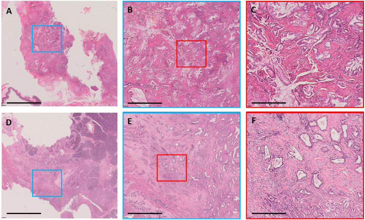
Figure 2.
Examples of stromal component. A case with low stromal component shown as whole-mount slide image in (A) (3.1 ×); the blue rectangular box was amplified in (B) (12.5×), and the red rectangular box was amplified in (C) (50×). A case with high stromal component shown as whole-mount slide image in (D) (3.1×); the blue rectangular box was amplified in (E) (12.5×), and the red rectangular box was amplified in (F) (50×). All images were stained with H&E. The black bar represents 8 mm in (A,D), 2 mm in (B,E), and 0.5 mm in (C,F).

