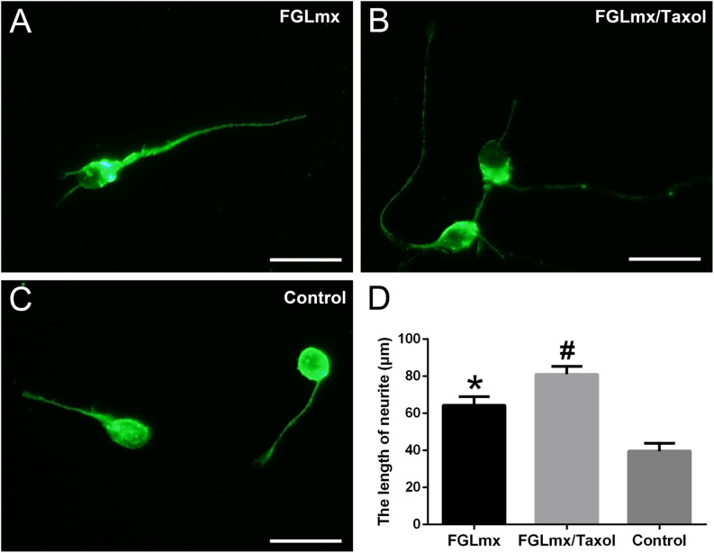
FIGURE 4.
The administration of Taxol released from FGLmx/Taxol increased neurite outgrowth from primary cortical neurons in vitro. βIII-Tubulin (green) staining of primary cortical neurons after 3 days of culture with (A) the supernatant of FGLmx, (B) supernatant of FGLmx/Taxol, or (C) PBS as a control. The scale bar represents 25 μm. (D) Quantification of the neurite length per well (± SEM). #P < 0.05 compared with the other two groups. *P < 0.05 compared with the control group.

