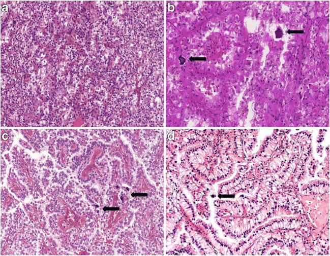
Fig. 1.
Representative images of typical morphological features of Xp11.2 renal cell carcinomas. (A) Solid-nested pattern with admixture of eosinophilic and clear cells. (B) Alveolar pattern populated by eosinophilic cells. Psammoma bodies are also present. (C) Papillary pattern with voluminous clear cells and psammoma bodies. D Occasionally the nuclei are near the apical surface of the cells and they mimic clear cell papillary renal cell carcinoma. The arrows indicate the psammoma bodies. All images have a magnification factor of 200x

