Figure 1.
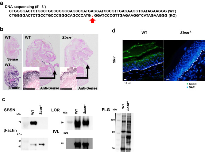
Characterization of Sbsn–/– mice. (a) DNA sequence. The red arrow indicates the mutation location. (b) Whole-mount in situ hybridization of E16.5. In WT mice, SBSN mRNA was markedly expressed in the dorsal skin, tail skin, and oral epithelium, but it was not seen in KO mice. (c) Western blotting. The samples were RIPA buffer extracts from the epidermis. SBSN was completely knocked out in Sbsn–/– mice. The expressions of LOR, IVL, and FLG were retained. The blots of SBSN and β-actin were cropped from one membrane. LOR, FLG, and IVL were cropped and reprobed from another membrane. (d) Immunofluorescence staining in footpad skin. Nuclei were counterstained with DAPI. SBSN (green) is observed in the upper epidermis and stratum corneum in WT, but not in Sbsn–/– mice. The lines illustrate the basal membrane of epidermis.

