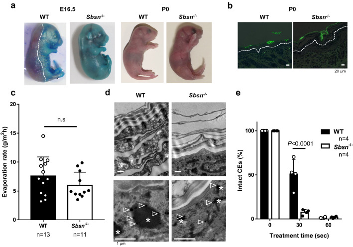
Figure 2.
Skin barrier function of Sbsn–/– mice. (a) Toluidine Blue staining. Left and right panels are E16.5 and newborn (P0), respectively. The line indicates the edge of the functional skin barrier. (b) Lucifer-Yellow penetration. Dorsal skin samples of the newborn were immersed in 1 mM lucifer yellow solution for 1 h. The lines indicate the basal membrane of epidermis. (c) Transepidermal water loss (TEWL). The inside-outside barrier function was assessed by TEWL. No difference is observed between WT and Sbsn–/– mice. (d) Ultrastructural analysis (TEM). Upper panel, stratum corneum; and lower panel, keratohyalin granules. *, keratohyalin granule; and arrowheads, ribosomes. (e) Fragility of cornified envelopes (CEs). CEs were sonicated for the indicated time. CEs isolated from Sbsn–/– mice were more rapidly destroyed by sonication than WT mice as indicated by the percentage of intact CEs.

