Figure 4.
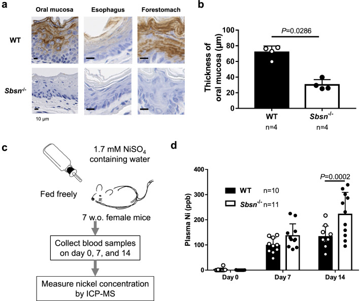
Blood nickel level in orally nickel-loaded Sbsn–/– mice. (a) Immunohistochemical staining in upper digestive tracts. Oral mucosa, esophagus, and forestomach were positively stained with anti-SBSN antibody in WT mice, but not in Sbsn–/– mice. Note that the epithelia of Sbsn–/– mice were thinner than those of WT mice. (b) Thickness of oral mucosa. The oral mucosa of Sbsn–/– mice was thinner than in WT. (c) Procedure of nickel loading test. The plasma nickel level was measured on day 0, 7, and 14. (d) Blood nickel level. The blood nickel level after oral feeding of nickel was higher in Sbsn–/– mice than in WT mice on day 14.

