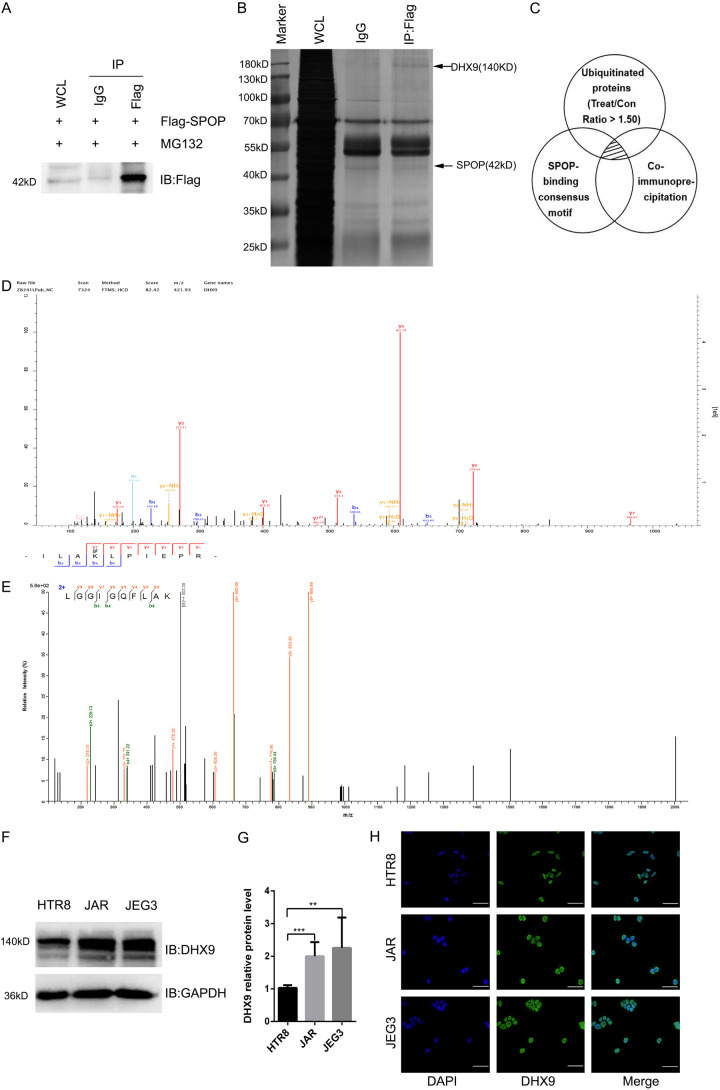
Figure 3.
DHX9 is low expressed in choriocarcinoma cells and is mainly localized in the nucleus. (A and B) IB analysis (A) and Silver stain assay (B) of JAR whole-cell lysates (WCLs) and anti-Flag immunoprecipitation derived from JAR cells transfected with the Flag-SPOP. The positions of DHX9 and SPOP were indicated by arrows. Rabbit immunoglobulin G (IgG) was used as a negative control for co-immunoprecipitations (co-IPs). Cells were treated with 10 μg/ml proteasome inhibitor MG132 for 10 h before harvesting. Flag, a tag protein. (C) Schematic diagram of three experimental methods. The shaded area indicates the proteins shared by the three experimental results. (D) The mass spectrum of DHX9 in ubiquitination experiment of JAR cells transfected with the indicated lentivirus. (E) The mass spectrum of DHX9 in co-immunoprecipitation assay of JAR cells transfected with the indicated lentivirus. (F and G) IB analysis of WCLs derived from HTR8, JAR and JEG3. Data are shown as mean ± SD of three independent experiments. **P < 0.01, ***P < 0.001, Student’s t-test. (H) Immunofluorescence for trophoblast cells HTR8, JAR and JEG3, cells were stained with DHX9 antibody (green), and the nuclei were counterstained with DAPI (blue). Scale bar, 50 um.

