Fig. 2.
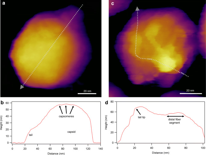
Imaging virions with atomic force microscopy (AFM) in non-contact mode, by using photothermal excitation to resonate the cantilever. a Height-contrast AFM image of a T7 bacteriophage particle attached to poly-l-lysine-coated mica. Based on the surface topography, this icosahedral virion is facing towards the buffer solution (phosphate-buffered saline, PBS) with its 3-fold symmetry axis. Individual capsomeres can be discerned in the image. b Topographical height profile plot along the axis of the particle image (indicated by the dotted straight line). c Height-contrast AFM image of a T7 bacteriophage particle pointing towards the buffer solution with its tail. To immobilize the tail fibers, the sample was chemically fixed with 2.5% glutaraldehyde and imaged in PBS. d Topographical height profile plot along the axis of a tail fiber (indicated by the dotted freehand line)

