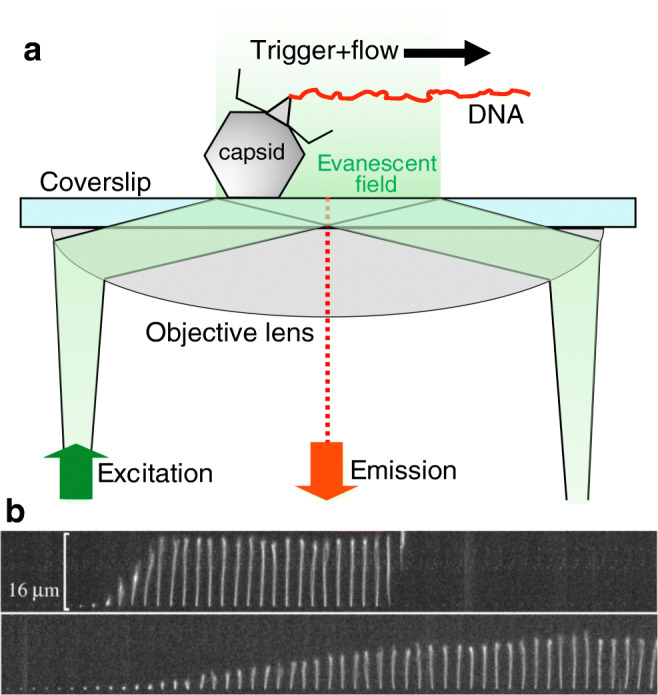
Fig. 6.

a Schematics of investigating the genomic DNA release from individual phage particles by using a microfluidic device, total internal reflection fluorescence (TIRF) microscopy, and DNA-intercalating fluorophores. b Time-resolved images of the release of dsDNA from a single λ-phage particle. DNA ejection was triggered by adding LamB (maltoporin), an E. coli outer-membrane protein, and the DNA molecule was visualized by rapid staining with the fluorescent dye SUBR Gold present in the buffer solution of the microfluidic chamber. Time delay between consecutive DNA images is 0.25 s. Upper and lower image series were recorded in the presence of 10 mM NaCl and 10 MgSO4, respectively. Adapted from (Grayson et al. 2007)
