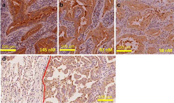
Fig. 7.
Each concentration 145 nM (a), 97 nM (b) and 58 nM (c) of mAb109 used as a primary antibody for stain the same human lung tumor samples. d Typical picture of IHC analysis of human lung tumor samples using mAb109 as primary antibody. (Red curve distinguishes the normal tissue (left) and adenocarcinoma (right))

