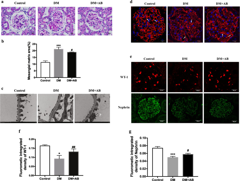
Fig. 5.
The effect of gut microbiota on renal injuries of incipient DN. a Pathological changes were assessed by PAS staining (original magnification, ×400). b The score of mesangial expansion was determined from histology sections. ***P < 0.01 compared with the control, #P < 0.05 compared with the DM group. c Changes in podocytes were evaluated by electron microscopy (original magnification, ×12 000). d The changes in glomerular endothelium glycocalyx were assessed by WGA staining (original magnification, ×400). e WT-1 and nephrin protein expression was evaluated by immunofluorescent staining (original magnification, ×400). The arrows indicate glomerular endothelium glycocalyx. f Quantitation of immunofluorescence staining for WT-1. *P < 0.05; compared with the control; ##P < 0.01 compared with DM. g Quantitation of immunofluorescence staining for nephrin. ***P < 0.001; compared with the control; #P < 0.05 compared with DM.

