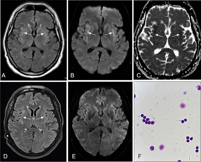
Fig. 1.
a-f MR scans 2 weeks (a–c) and 4 months (d–e) post symptom onset as well as CSF cytology (f): On axial FLAIR (a), Diffusion-weighted images (b) and the corresponding ADC map (c), bilateral signal alterations within the external capsule/claustrum regions are depicted (arrows), indicative of reduced diffusion. On follow-up imaging (d–e), the FLAIR-hyperintensities persist (d) whereas tissue diffusion has normalized (e). CSF-cytology (f) showed a slightly elevated cell count (9/µl) with a lymphocytic predominance (88% lymphocytes, 12% monocytes). A meaningful plasmacytic transformation was not observed, the monocytes being only slightly activated

