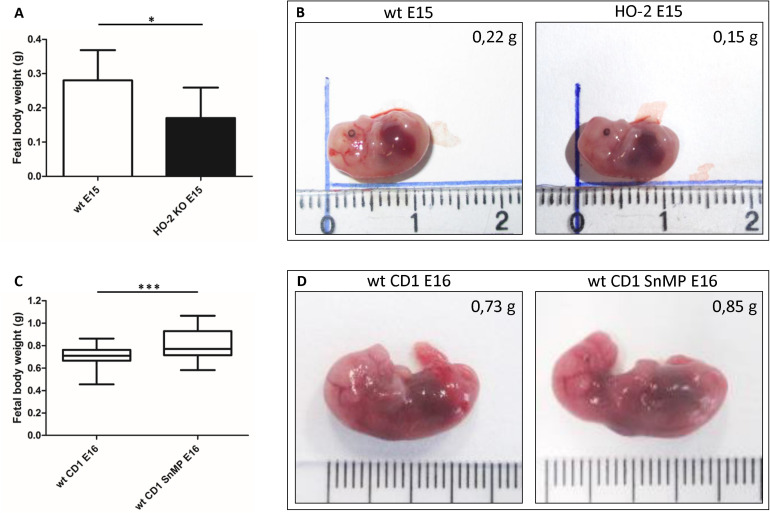
FIGURE 13.
Fetal body weight decreases by disruption of HO-2, but increases by HO-activity inhibition from E11. (A) Bar chart of the fetal body weight of the wt E15 fetuses (n = 15) and HO-2 KO E15 fetuses (n = 4), ∗p < 0.05. Data are shown as mean ± SD. (B) Representative fetus per group was shown (centimeter ruler). (C) Box-and-whisker plot with 10–90 percentiles of quantitative assessment of the body weight of the wt CD1 E16 fetuses (n = 91) and wt CD1 SnMP E16 fetuses (n = 56), ∗∗∗p < 0.001. (D) Representative fetus per group was shown (centimeter ruler).

