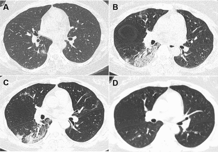
Fig. 1.
Dynamic changes of CT imaging in a 35-year-old woman with fever (37.8 °C) for 1 day. a At the first chest CT scan (day 1), a ground-glass lesion with partial consolidation was shown in the subpleural area of the right lower lobe; b at peak time (day 10), the lesion in the right lower lobe expanded with interlobular septal thickening and consolidation, and new lesions appeared in the upper lobes; c before discharge (day 17), the lesions were gradually absorbed and linear opacities were observed in the bilateral upper lobes; d after discharge (day 51), chest CT scan showed few GGO in the right lung, and the consolidation and linear opacities disappeared

