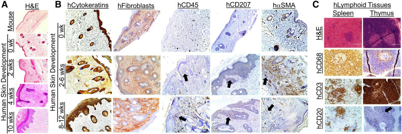
Figure 2.
Development of human skin and immune cells in the human Skin and Immune System-humanized NSG mouse model. (A) Representative histological (H&E) analysis of the human skin in human Skin and Immune System-humanized NSG mice (n = 4) demonstrate the development of human adult-like skin, including the dermis, multicellular layer (> 5 layers) epidermis, and cornified envelope. Representative histological (H&E) analysis of the mouse skin demonstrate a thin-layer of epidermal cells, with a thin dermal layer. (B) Various human skin cells are present in the human skin, including keratinocytes (AE1/AE3+ cells, hCytokeratins+ cells), dermal fibroblasts (TE7+ cells, hFibroblast+ cells), cutaneous immune cells (hCD45+ cells), and Langerhans cells (hCD207+); alpha-smooth muscle actin-expressing blood vessel cells (hα-SMA+ cells) are present in the human skin xenograft and expand during wound healing and contract after healing (n = 4). The black arrows denote representative IHC+ cells. (C) Representative histological and immunohistochemical analysis of the human spleen and thymus (both under the kidney capsule) in human Skin and Immune System-humanized NSG mice demonstrate the development of those lymphoid tissues at ten weeks post-transplantation, with human macrophages (hCD68+), T cells (hCD3+), B cells (hCD20+) present in the tissues (n = 4) Scale bars: 200 μm.

