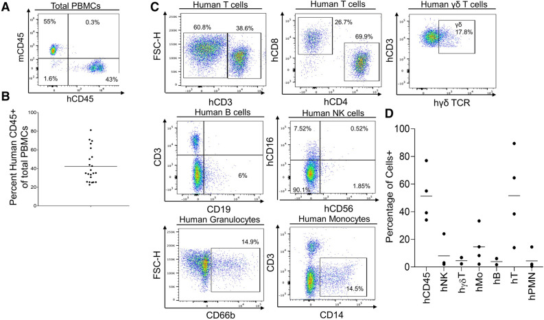
Figure 3.
Development of human peripheral blood mononuclear cells in the human Skin and Immune System-humanized NSG mouse model. (A) Representative flow cytometry analysis of human immune cell (hCD45+) reconstitution in peripheral blood mononuclear cells (PBMCs) of human Skin and Immune System-humanized NSG mice at 10–12 weeks post-transplantation demonstrates high levels (> 10%) of human immune cells in the blood. (B) Quantification of human immune cell reconstitution (n = 22; 3 independent experiments) in PBMCs of hSIS-humanized mice at 10 weeks post-transplantation. (C) Representative flow cytometry analysis of human PBMCs (hCD45+ PBMCs) in human Skin and Immune System-humanized mice at 10–12 weeks post-transplantation demonstrates readily detectable levels of various human immune cell types (B cells-hCD19+hCD3- human PBMCs, αβ T cells-hCD3+ human PBMCs, hCD4+ T cells, hCD8+ T cells, hγδTCR+ T cells- hγδ TCR+ CD3+ human PBMCs, natural killer cells (NK)-hCD57+ hCD3- human PBMCs, monocytes (hMo)-hCD14+ CD3- human PBMCs, and granulocytes (hPMN)-hCD66b+ hCD3- human PBMCs). (D) Quantification of human immune cell subtypes (n = 4) in human PBMCs (hCD45+ PBMCs) of human Skin and Immune System-humanized mice at 10–12 weeks post-transplantation.

