Figure 1.
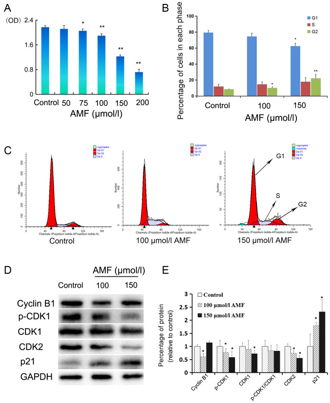
AMF decreases SKOV3 cell viability and induces cell cycle arrest. (A) SKOV3 cells were treated with different concentrations (0, 50 75, 100, 150 and 200 µmol/l) of AMF for 48 h and cell viability was assessed via the CellTiter 96 Aqueous One Solution Proliferation assay. The results demonstrated that AMF decreased SKOV3 cell viability in a dose-dependent manner. (B) Histograms showed the cell cycle distribution at G1, S and G2 phase. Data are presented as the mean ± SD (n=3). SKOV3 cells were treated with different concentrations of AMF (0, 100 and 150 µmol/l) for 48 h and cell cycle distribution was assessed via flow cytometric analysis. (C) Cell cycle analysis by flow cytometry. SKOV3 cells were treated with different concentrations of AMF (0, 100 and 150 µmol/l) for 48 h. (D) The expression levels of cyclin B, p-CDK1, CDK1, CDK2 and p21 were determined in SKOV3 cells treated with different concentrations of AMF (0, 100 and 150 µmol/l) for 48 h by western blot. GAPDH was used as the loading control. (E) Protein expression levels from the western blot in (D) relative to the GAPDH control. Data are presented as the mean ± SD (n=3). *P<0.05, **P<0.01 vs. control. AMF, amentoflavone; SD, standard deviation; CDK, cyclin-dependent kinase.

