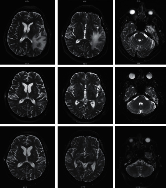
Figure 1.

T2-weighted sequences of the brain MRI demonstrating cerebral abscesses and associated edema. Top row: initial presentation; middle row: repeat imaging, 3 weeks after initial presentation; bottom row: final imaging, 2 months after initial presentation.
