Figure 6.

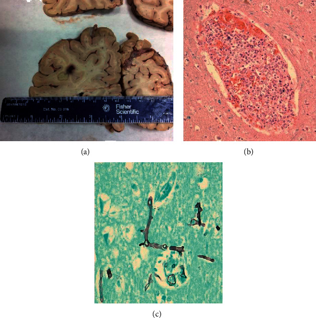
Autopsy: cerebral mucormycosis. Macroscopic appearance of temporal lobe abscess (a); abundant aseptate, wide-angled hyphae in blood vessels ((b), H&E stain) and brain parenchyma ((c), GMS stain).

Autopsy: cerebral mucormycosis. Macroscopic appearance of temporal lobe abscess (a); abundant aseptate, wide-angled hyphae in blood vessels ((b), H&E stain) and brain parenchyma ((c), GMS stain).