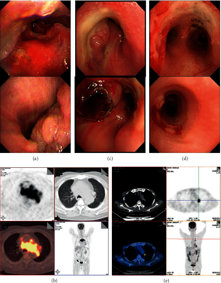
Figure 1.

Fiberoptic bronchoscopy and PET/CT images. (a) Fiberoptic bronchoscopy revealed intratracheal mass in right main bronchi with complete right mainstem bronchus occlusion before treatment. (b) PET/CT image showing left hilum and mediastinal mass (41 × 87 × 76 mm, FDG uptake 20.6) with compression of the pulmonary artery, lower tracheal segment, bilateral main bronchi, and superior vena cava. (c) Right mainstem bronchus was completely occluded, and a tracheal stent was implanted. (d) Bronchus occlusion was significantly improved after treatment with PD-1 inhibitor and DNMTi/HDACi. (e) Restaging PET image showing dramatically shrinkage of hilum and mediastinal mass (16 × 15 mm, FDG uptake 8.0) after a triple combination treatment of DNMTi/HDACi plus PD-1 inhibitor.
