
Figure 1.
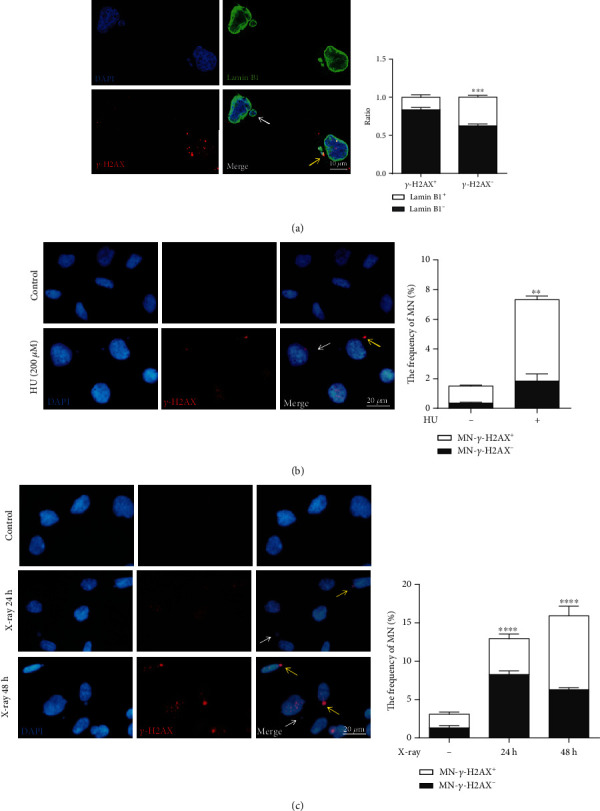
Micronuclei can be induced by genotoxicants. (a) Comparison of micronuclei membrane integrity between MN-γ-H2AX (+) and MN-γ-H2AX (-) in HT1080 cells. Representative images showing Lamin B1 (green) on the nuclear envelope, γ-H2AX (red) marking DNA double-strand break foci, and DAPI staining of DNA (blue). At least 200 MN of each type were counted. (b) The frequencies of MN measured by immunofluorescence in HT1080 cells treated with 200 μM HU for 24 h and washed out for 48 h. The left pictures show representative images. (c) The frequencies of MN in HT1080 cells at 24 h and 48 h after 10 Gy X-ray. The left pictures show representative images. Yellow arrows indicate MN-γ-H2AX (+); white arrows indicate MN-γ-H2AX (-). Each experiment was repeated three times. ∗∗P < 0.01, ∗∗∗P < 0.001, and ∗∗∗∗P < 0.0001. MN: micronuclei; HU: hydroxyurea.