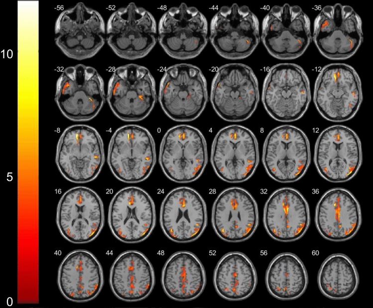
Figure 1.
Areas showing decreased GM volume in axial slices (p < 0.001) in Meige syndrome patients and control subjects. In the left hemisphere in the middle frontal orbital gyrus (t = − 8.83, p < 0.001), temporal pole superior temporal gyrus (t = − 8.20, p < 0.001) and insula (t = − 7.98, p < 0.001); in the right hemisphere in the temporal pole middle temporal gyrus (t = − 7.32, p < 0.001), precuneus (t = − 5.99, p < 0.001), inferior parietal (t = − 5.84, p < 0.001), inferior temporal (t = − 5.03, p < 0.001)and olfactory cortices (t = − 4.95, p < 0.001).

