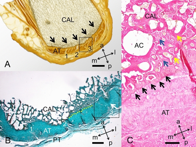
Figure 7.

The insertion of the Achilles tendon (AT) into the calcaneus (CAL) is depicted on plastinates and in histology. (A) Plastinated slice. Black arrows: demarcated trabeculae; dotted box: corresponds to an area similar to the dotted box in B; 1–3: fibrous septae, similar to the ones in B. Scale bar: 4 mm, (B) Masson–Goldner stain. Black box, median fibrous septum; PT, paratenon; 1–3: fibrous septae, similar to the ones in A; white arrow: median calcaneal prominence; yellow dotted line: demarcation between AT and CAL. Scale bar: 1 mm, (C) H&E stain. AC, adipocytes (located in bone marrow); black arrows: border between AT and CAL in greater detail compared to B; blue arrows: collagen bundles; white asterisk: folding artefact; yellow arrows: calcaneal trabeculae. Scale bar: 200 μm; a, anterior; l, lateral; m, medial; p, posterior.
