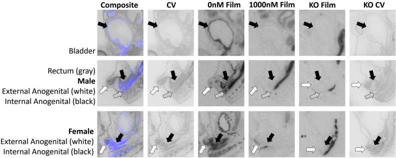
Figure 6.
AVPR1A ligand binding is evident in the genitourinary system and rectum of the neonatal mouse at P0. Arrows indicate region of interest. Composite = AVPR1A ligand binding and anatomy in a WT neonate (AVPR1A pseudo-colored in purple, cresyl violet counterstain in gray); CV = post-processed anatomical cresyl violet counterstain; 0 nM Film = AVPR1A ligand binding with no AVP competition in a WT neonate; 1000 nM Film = AVPR1A ligand binding in the 1000 nM AVP competition in a WT neonate; KO Film = AVPR1A ligand binding in AVPR1A KO mice; KO CV = post-processed anatomical cresyl violet stain of the AVPR1A KO neonate.

